Hepatectomy (Liver resection) for a rare case:
Giant liver hemangioma presenting as a cause of
fever of unknown origin
Dr. Prasad K. Wagle, Dr. Gunjan S. Desai
A 38-year-old lady, no co-morbidities Presentation: Fever of unknown origin (FUO). She was worked up exhaustively and meticulously for FUO, and infective, inflammatory, and autoimmune etiologies were ruled out.
During this period, her hemoglobin dropped from 12.6g/ dL to 9g/dL, without any overt signs of bleeding.
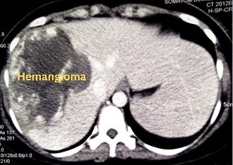
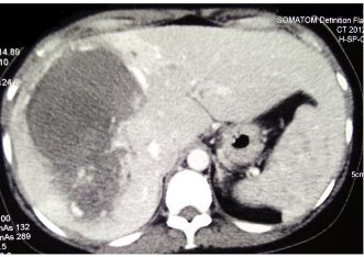
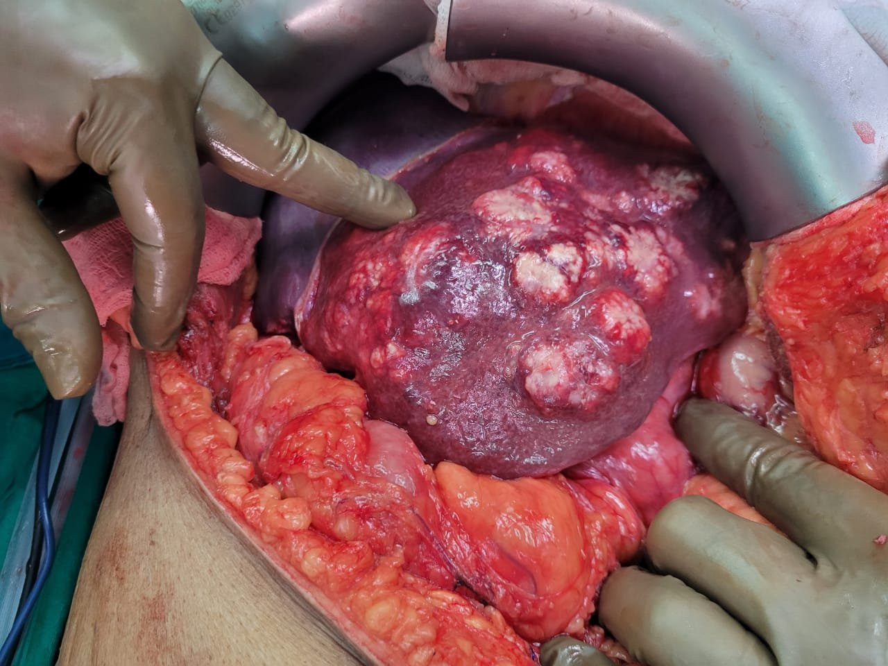
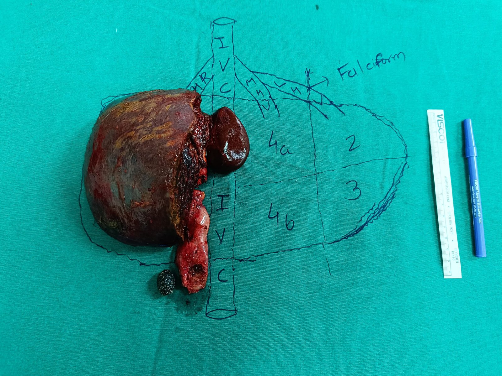
Abdominal ultrasonography(USG) / CECT scan – A large hemangioma involving hepatic segments 5, 6, 7, and 8 with areas of hemorrhage and thrombus in the lesion. Size – 15 x 12 x 8 cm PET scan – Given FUO, did not reveal any focus of infection.
Abdominal ultrasonography(USG) / CECT scan – A large hemangioma involving hepatic segments 5, 6, 7, and 8 with areas of hemorrhage and thrombus in the lesion. Size – 15 x 12 x 8 cm PET scan – Given FUO, did not reveal any focus of infection.
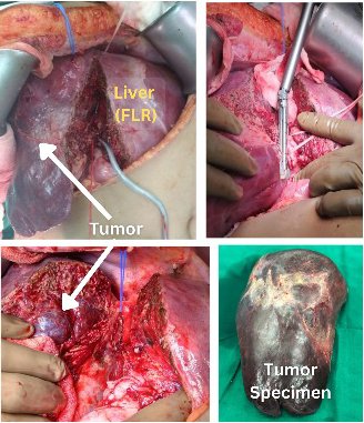
DIAGNOSIS: Giant Hepatic Hemangioma (A liver tumor) with hemorrhage and FUO Future liver remnant – 40% Bland arterial embolization was performed 24 hours before surgery.
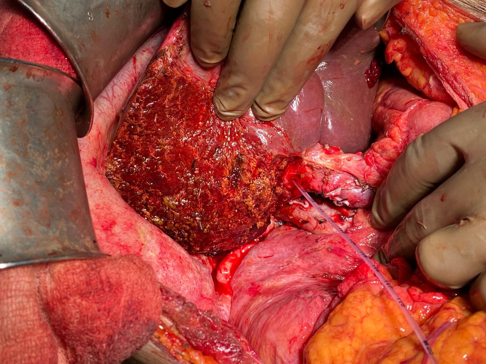
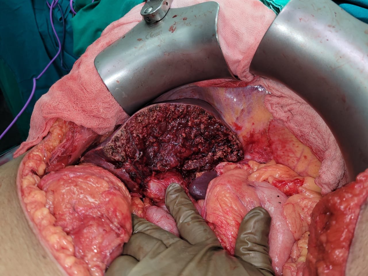
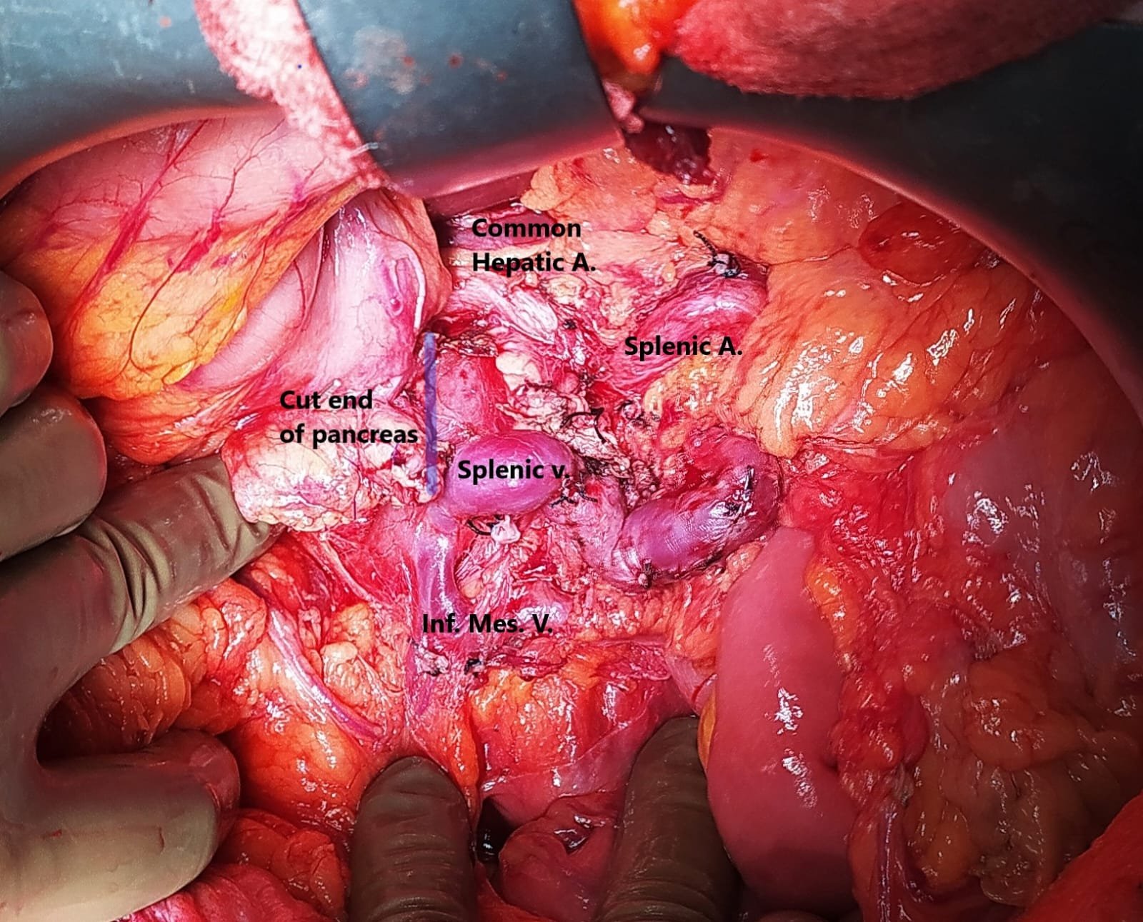
PLAN: RIGHT HEPATECTOMY
Post-operative recovery was uneventful.
Discharged on day 6 of surgery.
Fever subsided post-operatively.
HPE: Cavernous Hemangioma with areas of thrombosis and hemorrhage